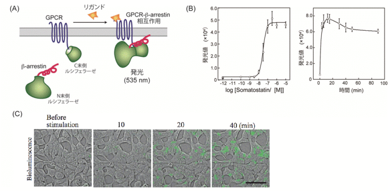
G protein-coupled receptors (GPCRs) are the most important target in drug discovery and are also important proteins for both basic and applied research. To understand the mechanism, we need to understand how GPCRs and other receptors work together. In order to understand this mechanism, we attempted to elucidate the mechanism of GPCRs by directly observing their intracellular movement.

Specifically, we added ligands and inhibitors to cells expressing formyl peptide receptor 1 (FPR1), one of the GPCRs, and observed Gα, a downstream molecule of FPR1, under a fluorescence microscope. For each of these states, we evaluated the time that Gα is bound to FPR1. The binding time of Gα was significantly longer only when the ligand was bound and the aggregates were formed. This result suggests that FPR1 strictly regulates signal transduction by functioning as an “AND gate,” a type of logic gate that produces an output when the two conditions of ligand binding and assembly are met. This observation was made by observing individual FPR1s on the plasma membrane while the cells were alive to detect the presence or absence of ligand binding and their assembly state. By detecting the three types of molecules (FPR1, ligand, and Gα) one by one in living cells and analyzing their motions and states, we were able to analyze the binding time of Gα for each of the four states of FPR1. The technique to analyze the dynamics of three types of molecules in a living cell simultaneously, one molecule at a time, is very rare in the world. If the state of activity and activation mechanism of each receptor can be elucidated, it is expected to provide useful knowledge not only for basic research but also for drug discovery and other applications.
- T. Nishiguchi, H. Yoshimura, R. S. Kasai, T. K. Fujiwara, T. Ozawa, ACS Chemical Biology. 15, 2577–2587 (2020).
