


Email: hideaki[at]chem.s.u-tokyo.ac.jp
略歴
| 平成13年 | 京都大学工学部工業化学科卒業 |
| 平成15年 | 京都大学大学院工学研究科分子工学専攻修士課程修了 |
| 平成19年 | 総合研究大学院大学物理科学研究科 構造分子化学専攻博士後期課程修了 博士(理学)取得 |
| 平成19年 | 京都大学再生医科学研究所 博士研究員 |
| 平成21年 | 東京大学大学院理学系研究科化学専攻 特任助教 |
| 平成27年 | 東京大学大学院理学系研究科化学専攻 助教 |
受賞等
| 2016 | ISPAC2016 Lecture Award (International Symposium on Pure and Applied Chemistry, Kuching, Malaysia) |
| 2015 | RSC Best Presentation Award (Royal Society of Chemistry Tokyo International Conference 2015) |
| 2012 | 日本化学会 第92回春季年会 優秀講演賞(学術) |
| 2006 | 日本化学会 第86回春季年会 学生講演賞 |
専門
- 生体分析化学
- 生物物理学
- 生体分子科学
主な研究テーマ
- 生細胞内分子の動態解析と操作を通じた生命現象作動機構の解明
- 主なターゲット:シグナル伝達、mRNA、機能性RNA
研究キーワード
1分子イメージング、光学顕微鏡、ライブイメージング、蛍光プローブ、生物発光、分子設計、分子操作、
RNA、シグナル伝達、生体分子集合体
Peer-Reviewed Papers
- Oligo DNA-based quantum dot (QD) single-particle tracking for multicolor single-molecule imaging
S. Sakuragi, N. Kato, T. Uchida, B. Zhao, T. Katagiri, M. Enomoto, R. Kato, H. Yoshimura, C. Oyama, I. Katayama, A. Chikuma, Y. Teramura, H. Bannai
Biophys. Pysicobiol., in press. DOI: 10.2142/biophysico.bppb-v23.0013 - Possible Direction of Drug Discovery Based on Single-Molecule Live Imaging
H. Yoshimura, T. Ozawa
Annu. Rev. Pharmacol. Toxicol., online ahead 66(1) 227-240 (2026) DOI: 10.1146/annurev-pharmtox-062624-025717 - Live-Cell Monitoring and Omics Analysis of Liquid-Solid Transitions of Biomolecular Condensates.
A. Farrag, K. Ota, H. Yoshimura, M. Takemoto, T. Mitarai, T. Kamikawa, M. Abo, V. Singh, C. Cui, L. Zhou, F. Ishidate, T. Fujiwara, S. Sato, Y. Hori, T. Ozawa, K. Kikuchi, and M. Uesugi*,
J. Am. Chem. Soc., 147(41) 37056–37064 (2025). DOI: 10.1021/jacs.5c07340 - Optical turbulence retrieval of heterogeneous media
M. Watabe, J. Sakamoto, H. Yoshimura, T. Nemoto, K. Kaizu
arXiv:2506.13204 (2025) DOI: 10.48550/arXiv.2506.13204 - Inducing aggresome and stable tau aggregation in Neuro2a cells with an optogenetic tool
S. Sakuragi, T. Uchida, N. Kato, B. Zhao, T. Takahashi, A. Hattori, Y. Sakata, Y. Soeda, A. Takashima, H. Yoshimura, G. Matsumoto, H. Bannai
Biophys. Physicobiol., 21(4) e210023 (2024). DOI: 10.2142/biophysico.bppb-v21.0023 - Intracellular tau fragment droplets serve as seeds for tau fibrils
Y. Soeda, H. Yoshimura, H. Bannai, R. Koike, I. Shiiba, A. Takashima
Structure, 32(19), 1793-1807.e6 (2024). DOI: 10.1016/j.str.2024.06.018 - Research on the molecular mechanism of singularity phenomenon in neurological disorders.
H. Bannai, A. Takashima, Y. Soeda, H. Yoshimura, G. Matsumoto, N. Sahara, M. Hiroshima, M. Hattori, T. Nagai,
Biophys. Physicobiol., e211008 (2024). DOI: 10.2142/biophysico.bppb-v21.s008 - Split Luciferase-Fragment Reconstitution for Unveiling RNA Localization and Dynamics in Live Cells.
M. Eguchi, H. Yoshimura, Y. Ueda, and T. Ozawa,
ACS Sensors, 8(11), 4055-4063 (2023). DOI: 10.1021/acssensors.3c01080 - Discovery of a phase-separating small molecule that selectively sequesters tubulin in cells.
G. Ado, N. Noda, H. T. Vu, A. D. Mahapatra, K. P. Arista, H. Yoshimura, D. M. Packwood, F. Ishidate, S. Sato, T. Ozawa, M. Uesugi,
Chem. Sci., 13, 5760-5766 (2022). DOI: 10.1039/d1sc07151c - Triple-color single-molecule imaging for analysis of the role of receptor oligomers in signal transduction.
H. Yoshimura*
* corresponding author
Biophys. Physicobiol., 19, 1-9 (2022). DOI: 10.2142/biophysico.bppb-v19.0007 - A Series of Furimazine Derivatives for Sustained Live-cell Bioluminescence Imaging and Application to the Monitoring of Myogenesis at Single-cell Level.
Mariko Orioka, Masatoshi Eguchi, Yuki Mizui, Yuma Ikeda Akihiro Sakama, Qiaojing Li, Hideaki Yoshimura*, Takeaki Ozawa, Daniel Citterio, Yuki Hiruta*
*co-corresponding author
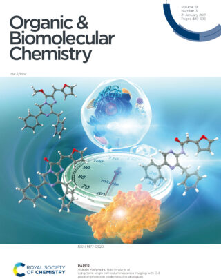
Bioconjugate Chem., 33, 496-504 (2022). DOI: 10.1021/acs.bioconjchem.2c00035 - Long-term single cell bioluminescence imaging with C-3 position protected coelenterazine analogues.
Y. Mizui, M. Eguchi, M. Tanaka, Y. Ikeda, H. Yoshimura*, T. Ozawa, D. Citterio, Y. Hiruta*
*co-corresponding author
Org. Biomol. Chem., 19, 579-586 (2021). DOI: 10.1039/d0ob02020f - Potential of Single-Molecule Live-Cell Imaging for Chemical Translational Biology.
H Yoshimura*
*corresponding author
ChemBioChem, 22, 2941-2945 (2021). DOI: 10.1002/cbic.202100258 - Signaling activations through G-protein-coupled-receptor aggragations.
M. Watabe, H. Yoshimura, S. N. V. Arjunan, K. Kaizu, K. Takahashi
Phys. Rev. E, 102, 032413 (2020). DOI:10.1103/PhysRevE.102.032413 - Synergetic roles of Formyl Peptide Receptor 1 oligomerization in ligand-induced signal transduction.
T. Nishiguchi, H. Yoshimura, R.S. Kasai, T. K. Fujiwara, T. Ozawa
ACS Chem. Biol., 15, 2577-2587 (2020). DOI: 10.1021/acschembio.0c00631 - Photocleavable Cadherin Inhibits Cell-to-Cell Mechanotransduction by Light.
M. Endo, T. Iwawaki, H. Yoshimura, and T. Ozawa
ACS Chem. Biol., 14, 2206-2214 (2019). DOI: 10.1021/acschembio.9b00460 - A Robust Split-Luciferase-Based Cell Fusion Screening for Discovering Myogenesis-Promoting Molecules
Q. Li, H. Yoshimura, M. Komiya, K. Tajiri, M. Uesugi, Y. Hata, T. Ozawa
Analyst, 143, 3472-3480 (2018). DOI: 10.1039/C8AN00285A - Unique Roles of β-Arrestin in GPCR Trafficking Revealed by Photoinducible Dimerizers.
O. Takenouchi, H. Yoshimura, T. Ozawa
Sci. Rep., 8, 677 (2018). DOI: 10.1038/s41598-017-19130-y - Protein dynamics of the oxygen sensor protein HemAT as revealed by time-resolved step-scan FTIR spectroscopy.
A. Pavlou, H. Yoshimura, S. Aono, E. Pinakoulaki
Biophys. J., 114, 584-591 (2018). DOI: 10.1016/j.bpj.2017.12.012 - Live Cell Imaging of Endogenous RNAs Using Pumilio Homology Domain Mutants: Principles and Applications.
H. Yoshimura*
*corresponding author
Biochemistry, 57, 200-208 (2018). DOI: 10.1021/acs.biochem.7b00983 - Real-time fluorescence imaging of single-molecule endogenous non-coding
RNA in living cells.
H. Yoshimura and T. Ozawa,
Methods Mol. Biol., 1649, 337-347 (2018).DOI: 10.1007/978-1-4939-7213-5_22 - Probing the role of the heme distal and proximal environment in ligand dynamics in the signal transducer protein HemAT by time-resolved step-scan FTIR and resonance Raman spectroscopy.
A. Pavlou, A. Loullis, H. Yoshimura, S. Aono, E. Pinakoulaki,
Biochemistry, 56, 5309-5317 (2017). DOI: 10.1021/acs.biochem.7b00558 - Spatiotemporal analysis with a genetically encoded fluorescent RNA probe reveals TERRA function around telomeres.
T. Yamada, H. Yoshimura, R. Shimada, M. Hattori, M. Eguchi, T. K. Fujiwara, A. Kusumi, T. Ozawa,
Sci. Rep. 6, 38910 (2016). DOI: 10.1038/srep38910 - Monitoring of RNA dynamics in living cells using PUM-HD and fluorescent protein reconstitution technique.
H. Yoshimura and T. Ozawa,
Methods Enzymol., 572, 65-85 (2016). DOI: 10.1016/bs.mie.2016.03.018 - Genetically Encoded Fluorescent Probe for Imaging Apoptosis in Vivo with Spontaneous GFP Complementation.
Y. Nasu, Y. Asaoka, M. Namae, H. Nishina, H. Yoshimura, T. Ozawa,
Anal. Chem., ;88, 838-844 (2016). DOI: 10.1021/acs.analchem.5b03367 - Development of red-shifted mutants derived from luciferase of Brazilian click beetle Pyrearinus termitilluminans
T. Nishiguchi, T. Yamada, Y. Nasu, M. Ito, H. Yoshimura, T. Ozawa
J. Biomed. Opt.,20, 101205 (2015).DOI: 10.1117/1.jbo.20.10.101205 - Simultaneous time-lamination imaging of protein association using a split fluorescent timer protein.
A. Takamura, M Hattori, H. Yoshimura, T Ozawa
Anal. Chem., 87, 3366-3372 (2015). DOI: 10.1021/ac504583t - Method of split-reporter reconstitution for the analysis of biomolecules
H. Yoshimura, T. Ozawa
Chem. Rec, 14, 492-501 (2014). DOI: 10.1002/tcr.201402001 - Bioluminescent Probes to Analyze Ligand-induced Phosphatidylinositol
3,4,5-trisphosphate Production with Split Luciferase Complementation
L.Z. Yang, Y. Nasu, M. Hattori, H. Yoshimura, A. Kanno, T. Ozawa,
Anal. Chem., 85, 11352-11359 (2013). DOI: 10.1021/ac402278f - Advances in fluorescence and bioluminescence imaging
T. Ozawa, H. Yoshimura and S.B. Kim,
Anal. Chem.,85, 590-609 (2013). DOI: 10.1021/ac3031724 - Fluorescent probes for imaging endogenous β-actin mRNA in living cells using fluorescent protein-tagged pumilio.
H. Yoshimura, A. Inaguma, T. Yamada and T. Ozawa
ACS Chem. Biol., 7, 999-1005 (2012). DOI: 10.1021/cb200474a - Visualization of non-engineered single mRNAs in living cells using genetically encoded fluorescent probes.
T. Yamada, H. Yoshimura, A. Inaguma and T. Ozawa,
Anal. Chem., 83, 5708-5714 (2011). DOI: 10.1021/ac2009405 - Hydrogen bonding interaction on the heme-bound ligand in the heme-based O2 sensor protein,
M. Nishimura, H. Yoshimura, K. Ozawa, S. Yoshioka, M. Kubo, T. Kitagawa and S. Aono,
J. Porphyrins Phthalocyanines, 12, 142-148 (2008). DOI: 10.1142/S1088424608000182 - Protein conformation changes of HemAT-Bs upon ligand binding probed by ultraviolet resonance Raman spectroscopy.
S. F. EI-Mashtoly, Y. Gu, H. Yoshimura, S. Yoshioka, S. Aono, T. Kitagawa,
J. Biol. Chem., 283, 6942-6946 (2008). DOI: 10.1074/jbc.M709209200 - The signal transduction mechanism of HemAT-Bs through the proximal heme pocket revealed by time-resolved resonance Raman spectroscopy.
H. Yoshimura, S. Yoshioka, Y. Mizutani and S. Aono,
Biochem. Biophys. Res. Commun., 307, 1053-1057 (2007). DOI: 10.1016/j.bbrc.2007.04.041 - Two ligand binding sites in the O2-sensing signal transducer HemAT: implications for ligand recognition/discrimination and signaling,
E. Pinakoulaki, H. Yoshimura, V. Daskalakis, S. Yoshioka, S. Aono and C. Varotsis,
Proc. Natl. Acad. Sci. USA, 103, 14796-14801 (2006). DOI: 10.1073/pnas.0604248103 - Specific hydrogen-bonding networks responsible for selective O2 sensing of the oxygen sensor protein HemAT from Bacillus subtilis,
H. Yoshimura, S. Yoshioka, K. Kobayashi, T. Ohta, T. Uchida, M. Kubo, T. Kitagawa and S. Aono,
Biochemistry, 45, 8301-8307 (2006). DOI: 10.1021/bi060315c - Recognition and discrimination of gases by the oxygen-sensing signal transducer protein HemAT as revealed by FTIR spectroscopy,
E. Pinakoulaki, H. Yoshimura, S. Yoshioka, S. Aono and C. Varotsis,
Biochemistry, 45, 7763-7766 (2006). DOI: 10.1021/bi0604072 - Non-covalent modification of the heme-pocket of apomyoglobin by a 1,10-phenanthroline derivative,
Y. Hitomi, H. Mukai, H. Yoshimura, T. Tanaka and T. Funabiki,
Bioorg. Med. Chem. Lett., 16, 248-251 (2006). DOI: 10.1016/j.bmcl.2005.10.016 - Biophysical properties of a c-type heme in chemotaxis signal transducer protein DcrA,
S. Yoshioka, K. Kobayashi, H. Yoshimura, T. Uchida, T. Kitagawa and S. Aono,
Biochemistry, 44, 15406.-15413 (2005). DOI: 10.1021/bi0513352 - Oxygen-sensing mechanism of HemAT from Bacillus subtilis: a resonance Raman spectroscopic study,
T. Ohta, H. Yoshimura, S. Yoshioka, S. Aono and Teizo Kitagawa,
J. Am. Chem. Soc., 126, 15000-15001 (2004). DOI: 10.1021/ja046896f
著書・総説・解説
- 論文図表を読む作法PREMIUM
吉村英哲 (担当:分担執筆, 範囲:第3章 イメージング 11 全反射照明蛍光顕微鏡)
羊土社 2025年12月11日 (ISBN: 9784758122863) - シグナル伝達を精密に調節する細胞内の分子機械
吉村英哲
「現代化学」(東京化学同人)No. 651 (2025年6月号) p78 「論文ファイルから」 - オプトバイオロジー
吉村英哲、小澤岳昌
「未来技術 2025-2034 全産業編」(日経BP)第16章 8節 - A Method for Bioluminescence-Based RNA Monitoring Using Split-Luciferase Reconstitution Techniques
Masatoshi Eguchi, Hideaki Yoshimura, Takeaki Ozawa
Methods Mol. Biol., 2875, 9-20 (2025) - mRNA可視化プローブの理論
吉村英哲、小澤岳昌
「mRNAの制御機構の解明と治療薬・ワクチンへの活用」(技術情報協会刊)第8節 - Optical monitoring of single molecule dynamics of RNA in living cells
Hideaki Yoshimura*, Takeaki Ozawa
* corresponding author
Springer Series in Chemical Physics,”Progress in Photon Science”, 95-106 (2021) - 蛍光イメージング分光法(FRET, FCS, FCCS):生細胞への応用
吉村英哲
日本分光学会監修 『紫外可視・蛍光分光法』(講談社サイエンティフィク刊) 第3章, (2021) - 蛍光プローブに用いられる色素の一覧
吉村英哲
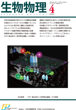
日本分光学会監修 『紫外可視・蛍光分光法』(講談社サイエンティフィク刊) 付録, (2021) - 細胞膜受容体の集合とシグナル伝達の3色同時蛍光1分子イメージングによる解析
吉村英哲
*corresponding author
生物物理, 61(4), 245-247 (2021). DOI: 10.2142/biophys.61.245 - A Split-Luciferase-Based Cell Fusion Assay for Evaluating the Myogenesis-Promoting Effects of Bioactive Molecules
Q. Li, H Yoshimura*, T. Ozawa
* corresponding author
Methods in Mol. Biol., 2274, 79-87 (2021). DOI: 10.1007/978-1-0716-1258-3_8 - Quantitative Analysis of Membrane Receptor Trafficking Manipulated by Optogenetic Tools
O. Takenouchi, H Yoshimura*, T. Ozawa
* corresponding author
Methods in Mol. Biol., 2274, 15-23 (2021). DOI: 10.1007/978-1-0716-1258-3_2 - 二分割ルシフェラーゼ再構成法による発光プローブ
吉村英哲、小澤岳昌
実験医学別冊「発光イメージング実験ガイド」(羊土社刊)プロトコール編 II-4 p62-70, 2019 - 細胞膜は二次元流体の夢を見るか?
吉村英哲
月刊「化学」(化学同人社刊), Vol 74, No. 5, pp70-71,2019 - Optical Control of G Protein-Coupled Receptor Activated in Living Cells
Hideaki Yoshimura, Takeaki Ozawa
Springer Series in Chemical Physics,”Progress in Photon Science: Recent Advances”, Chapter 7, pp.129-138, Springer (2018). DOI: 10.1007/978-3-030-05974-3_7 - 生命分子の機能を超えるための解析化学
吉村英哲、小澤岳昌
CSJ Current Review 30 生命機能に迫る分子科学:生命分子を真似る、飾る、超える(化学同人社刊)第2章 p28-33, 2018 - 蛍光顕微鏡を用いた生細胞内1分子可視化解析法
吉村英哲、小澤岳昌
ナノバイオ・メディシン 細胞核内反応とゲノム編集(近代科学社刊)第2章 p32-45, 2017 - RNAと生細胞内1分子イメージングの可能性
吉村英哲
Labcab p19-20, Vol.12 No.1 2015 - 蛍光顕微鏡を用いた生細胞内1分子可視化解析法
吉村英哲、小澤岳昌
The Bulletin of the Society of Nano Science and Technology (ナノ学会会報)
第13巻 第2号 2015年3月,pp61-65
ISSN 1347-8028 - 生細胞内RNAイメージング
吉村英哲、小澤岳昌
細胞工学 (秀潤社) Vol.34, No.1, pp53-58, 2015 - RNAイメージング
吉村英哲
東京大学理学系研究科・理学部ニュース 連載:理学のキーワード
Vol.44, No.5, pp12-13, 2013
招待講演・依頼講演
- TBA
分析化学会 第75年会 2026年9月15日 - TBA
日本膜学会 第48年会 2026年6月1日 - “Time-consecutive single-molecule live-cell imaging to analyze the receptor cluster formation process at the plasma membrane”
2nd International Symposium on Chemical & Biomedical Imaging (2026年5月28日 The Hong Kong Polytechnic University) - “Time-consecutive molecular-motion mapping visualizes inhomogeneous motion of molecules in living cells”
18th International Symposyum on Nanomedicine (2025年12月3日 広島大学) - “Observation and analysis of single-molecule motility in living cells through cell physiological events”
OPIC2025 –Optics & Photonics International Congress (2025年 4月 23日 パシフィコ横浜) - “分子運動マッピングによる生細胞内シグナル伝達機構の可視化”
第24回生命化学研究会ポストコンファレンス(2024年12月16日) - “Large-scale live-cell single-molecule imaging to monitor spatially and temporally inhomogeneous molecular motion”
17th Internationaly Symposium on Nanomedicine(2024年12月3日 名古屋工業大学) - “生命の分子機構解明を目指す生細胞内大規模1分子追跡に基づくアプローチ”
第97回日本生化学会大会(2024年11月6日 パシフィコ横浜) - “細胞内分子運動の1分子観察で見えてくること”
第7回分子ロボティクス年次大会(2024年3月13日 東京大学) - “蛍光と生物発光で見る生細胞内分子動態”
2023年度日本分光学会北海道支部シンポジウム(2024年2月22日 北海道大学) - “Monitoring RNA Motility in Living Cells Using Fluorescence and Bioluminescence”
16th International Symposium on Nanomedicine (2023年11月21日 大阪公立大学杉本キャンパス) - “Analysis of molecular motility in living cells for understanding mechanisms of living system through single-molecule imaging”
15th International Symposium on Nanomedicine (2022年12月6日 徳島大学・長井記念ホール) - “生細胞RNAイメージング技術とその可能性”
第45回日本分子生物学会年会(2022年11月30日 幕張メッセ) - “細胞内分子動態の蛍光1分子観察でわかること”
第20回 関東光科学若手研究会(2022年11月5日 東京大学) - “生細胞1分子イメージングで見るRNAの動態と作動機構”
第4回 形態解析ワークショップ(2022年5月7日 順天堂大学) - “Oligomer Formation and Signal Transduction of GPCR -A Single-Molecule Live Imaging Study”
14th International Symposium on Nanomedicine, Taiwan-Japan-Korea Nanomedicine Meeting (2021年11月19日 online) - “蛍光イメージング分光法 − 生細胞への応用と蛍光ライブイメージング”
光とレーザーの科学技術フェア2021 (2020年11月17日 東京都立産業貿易センター浜松町館) - “生命の仕組みを解明する蛍光分析 -生きたままの細胞を対象に-”
光とレーザーの科学技術フェア2020 (2020年11月11日 東京都立産業貿易センター浜松町館) - “The roles of receptor oligomerization for signal transduction - A study through single-molecule live-cell imaging-“
13th International Symposium on Nanomedicine (2019年12月5日 甲南大学ポートアイランドキャンパス) - “分子動態からメカニズムを探る -生細胞1分子イメージングを用いたアプローチ-“
第11回光塾 (2019年11月13日 理化学研究所神戸キャンパス) - “Novel optical techniques to explore biological function in single cells”
The 4th STEPS Symposium on Photon Science (2019年3月21日 東京大学本郷キャンパス) - “分子動態解析による細胞内分子作動機構の解明-生細胞1分子イメージングによるアプローチ-“
第95回創薬科学セミナー (2019年2月22日 名古屋大学) - “A single molecule imaging approach to understand signal transduction on the plasma membrane in living cells”
12th International Symposium on Nanomedicine (2018年12月7日 山口大学小串キャンパス) - “Simultaneous single molecule observation of telomeric-repeat containing RNA and proteins in living cells”
CMCB2017 (2017年4月26日 Hilton Xi’an, China) - “Simultaneous single molecule imaging of non-coding RNA and proteins in living cells”
AnalytiX 2017 (2017年3月24日 福岡) - “Single molecule imaging in living cells to reveal the relationship between motions and functions of biological molecules”
10th International Symposium on Nanomedicine(2016年11月24日 産業技術総合研究所) - “Single molecule imaging approach to reveal molecular motions and functions in cellular events”
1st Nano/Bioscience International Syposium (2016年10月7日 同志社大学) - “Tailor-made design of protein-based probes to visualize and analyze single-molecule motion of RNAs in living cells “
International Symposium on Pure and Applied Chemistry 2016 (ISPAC2016)
(2016年8月16日 Borneo Convention Center Kuching, Malaysia) - “Analysis of distribution and motion of a non-coding RNA in living cells using a single molecule tracking approach”
The 5th Serendipiter seminar (JST-ImPACT program)
(2016年7月26日 東京大学) - “Single molecule imaging of telomeric-repeat containing RNA in living cells”
9th International Symposium on Nanomedicine(2015年12月12日 三重大学) - “Single molecule imaging to reveal mechanisms of complex cellular systems”
NTU-SNU-UT Chemistry Symposium(2015年1月16日 National Taiwan University) - “Single molecule imaging in living cells to reveal the relationship between motions and functions of biological molecules”
8th International Symposium on Nanomedicine(2014年12月6日 愛媛大学) - “A study of a molecular mechanism of intracellular signal transduction based on single-molecule imaging”
The 2nd Japan-China Symposium on Nanomedicine(2014年5月17日 広島大学霞キャンパス) - “Analysis of RNA dynamics in living cells based on single molecule imaging”
7th International Symposium on Nanomedicine(2013年11月9日 九州工業大学) - “Visualization of molecular motion in living cells using a single molecule imaging method”
錯体化学会第63回討論会(2013年11月3日 琉球大学千原キャンパス)
アウトリーチ活動
- 第18回分析化学会近畿支部夏季セミナー 講師
演題:”アカデミアで生きていく”
2024年8月5日 SORA RINKU - 第5回 化学発光イメージングワークショップ 実習講師
2020年 2月19日−20日 東京大学理学部化学館 - The 1st International Training Course for Singularity Biology, Lecturer
演題:”Bioiluminescence probes to detect and analyze singularity events”
2019年8月5日 大阪大学 - 第4回 化学発光イメージングワークショップ 実習講師
2019年 1月29日−30日 大阪大学産業科学研究所 - 東京大学オープンキャンパス 理学部化学科講演会 講師
演題:「分子のサイズで生き物を観てみると」
2018年8月1日 東京大学理学部化学講堂 - 第2回化学発光イメージングワークショップ 実習講師
2017年2月8日−9日 大阪大学産業化学研究所 - UTalk 講師
演題:「色づけしてみた生命」
2013年12月14日 東京大学情報学環・福武ホール - 日本化学会 第94春季年会 分析化学ディビジョンプログラム編成委員
2013年8月〜12月
学会活動・委員等
- 第11回 バイオ関連化学シンポジウム若手フォーラム 代表世話人
2024年9月11日開催 - 日本生物物理学会 分野別専門委員
2023年1月〜2023年12月 - 第45回日本分子生物学会年会ワークショップ オーガナイザー
「分子を観る・分子で観る:ケミカルバイオイメージングの新展開」 - 日本化学会バイオテクノロジー部会 運営委員
2022年9月〜 - 「光塾」総括塾員
2019年11月〜 - 日本化学会 第94春季年会 分析化学ディビジョンプログラム編成委員
2013年8月〜12月
競争的研究費獲得状況
科学研究費補助金
- 挑戦的研究(開拓) (代表)
「細胞試料を生かしたまま1分子4D観察する革新的バイオイメージング技術の創出」
2023年6月-2026年3月
総額26,000千円 (直接経費20,000千円) - 挑戦的研究(萌芽) (分担; 代表者 坂内博子 早稲田大学教授)
「1分子計測によるタウオパチー発症の開始点と多様性の起源の研究」
2023年6月-2025年3月
総額1,300千円 (直接経費1,000千円) - 基盤研究(B) (代表)
「分子システムとして細胞機能を理解する時空間拡大型生細胞1分子イメージング法の創出」
2023年4月-2026年3月
総額19,240千円 (直接経費14,800千円) - 挑戦的研究(開拓) (代表)
「遺伝子発現の時空間可視化追跡を生体試料を生かしたまま実現する技術群の創出」
2020年7月-2023年3月
総額26,000千円 (直接経費20,000千円) - 基盤研究(B) (代表)
「RNA可視化法と発光イメージング法に基づく生細胞遺伝子発現時空間解析法の創出」
2019年4月-2022年3月
総額17,680千円 (直接経費13,600千円) - 新学術領域研究(研究領域提案型) (分担;代表者 永井健治 大阪大学栄誉教授)
「シンギュラリティ細胞を探索・操作するための細胞機能3次元可視か・光操作技術の開発」
2019年6月-2023年3月
総額33,540千円 (直接経費25,800千円) - 基盤研究(B) (代表)
「生細胞内RNA動態の包括的可視化分析技術の創発」
2016年4月-2019年3月
総額18,070千円 (直接経費13,900千円) - 挑戦的萌芽研究 (代表)
「1分子レベルでの生細胞内RNAリアルタイム計数プローブの開発」
2014年4月-2016年3月
総額4,030千円 (直接経費3,100千円) - 若手研究(A) (代表)
「生細胞内RNAの可視化と制御を実現する革新的技術の創出」
2013年4月-2016年3月
総額26,260千円 (直接経費20,200千円) - 若手研究(B) (代表)
「受容体クラスター形成の光制御による細胞への人工的生態シグナル入力法の開発」
2011年4月-2013年3月
総額4,680千円 (直接経費3,600千円)
科研費以外の研究費
- 旭硝子財団 ステップアップ助成 (代表)
「分子から多細胞をシームレスにつなぎ生体機能を誘起する分子運動を解析する統合技術群の創出」
2024年4月-2027年3月
総額14,000千円 (直接経費14,000千円) - 旭硝子財団 若手継続グラント (代表)
「多細胞サンプル内における遺伝子発現1細胞長時間定量追跡法の開発」
2019年4月-2022年3月
総額6,000千円 (直接経費6,000千円) - 2019年度 物質・デバイス領域共同研究拠点 基盤共同研究課題 (代表)
「生物発光イメージングによる生細胞内遺伝子発現可視化追跡法の創出」
2019年4月-2020年3月
総額100千円 (直接経費100千円) - 山田科学振興財団 研究助成 (代表)
「1分子動態検出に基づく細胞内シグナル伝達量測定法の開発」
2018年10月-2020年3月
総額2,000千円 (直接経費2,000千円) - 平成30年トヨタ理研スカラー (代表)
「mRNA機能の生細胞内空間特異的操作法の創出」
2018年4月-2019年3月
総額1,000千円 (直接経費1,000千円) - 内藤記念科学振興財団 内藤記念特定研究助成金 (代表)
「機能性RNA TERRAの生細胞内1分子イメージングによる機能発現機構の解明」
2017年12月-
総額500千円 (直接経費500千円) - 旭硝子財団 研究奨励(代表)
「1細胞内RNA定量経時追跡法の開発」
2016年4月-2018年3月
総額2,000千円 (直接経費2,000千円) - 住友財団 基礎科学研究助成(代表)
「非翻訳性RNAによるテロメア長調節機構の解明−生細胞内1分子動態解析による研究」
2015年11月-2017年11月
総額1,800千円 (直接経費1,800千円) - 日本科学協会 笹川科学研究助成 (代表)
「生細胞内RNAのリアルタイム計数および1分子動態追跡法の開発」
2014年4月-2015年2月
総額800千円 (直接経費800千円) - 平成22年度GCOE若手助成補助金 (代表)
「光制御を利用した空間勾配のある生体シグナル細胞への導入法の開発」
2010年8月-2011年4月
総額800千円 (直接経費800千円)